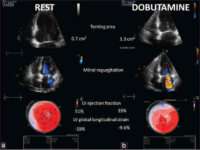
Dobutamine Stress Echocardiogram
An echocardiogram (echo) is a test used to assess the heart's function and structures. A stress echocardiogram is a test done to assess how well the heart works under stress. The “stress” can be triggered by either exercise on a treadmill or medication called dobutamine.
A dobutamine stress echocardiogram (DSE) may be used if you are unable to exercise. Dobutamine is put in a vein and causes the heart to beat faster. It mimics the effects of exercise on the heart.
During an echo, a transducer (like a microphone) sends out ultrasonic sound waves at a frequency too high to be heard. When the transducer is placed on the chest at certain locations and angles, the ultrasonic sound waves move through the skin and other body tissues to the heart tissues, where the waves bounce or "echo" off of the heart structures. The transducer picks up the reflected waves and sends them to a computer. The computer displays the echoes as images of the heart walls and valves.
A DSE may involve one or more of these special types of echocardiograms:
- M-mode echocardiogram. This, the simplest type of echocardiogram, produces an image that is similar to a tracing rather than an actual picture of heart structures. M-mode echo is useful for measuring heart structures, such as the heart's pumping chambers, the size of the heart itself, and the thickness of the heart walls.
- Doppler echocardiogram. This Doppler technique is used to measure and assess the flow of blood through the heart's chambers and valves. The amount of blood pumped out with each beat is a sign of how well the heart is working. Also, Doppler can detect abnormal blood flow within the heart, which can mean there is a problem with one or more of the heart's four valves or with the heart's walls.
- Color Doppler. Color Doppler is an enhanced form of a Doppler echocardiogram. With color Doppler, different colors are used to show the direction of blood flow.
- 2-D (two-dimensional) echocardiogram. This technique is used to see the actual structures and motion of the heart structures. A 2-D echo view looks cone-shaped on the monitor, and the real-time motion of the heart's structures can be seen. This allows the doctor to see the various heart structures at work and evaluate them.
- 3-D (three-dimensional) echocardiogram. 3-D echo technique captures 3-D views of the heart structures with greater depth than 2-D echo. The live or "real time" images allow for a more accurate assessment of heart function by using measurements taken while the heart is beating. 3-D echo shows enhanced views of the heart's anatomy and can be used to determine best treatment plan.
